 The knee is one of the joints most prone to injury. Its structure and many components put it at risk of many types of injuries, which can result in knee pain or loss of function. Injuries of the muscles and tendons surrounding the knee are caused by acute hyperflexion or hyperextension of the knee or by overuse. These injuries are called strains.
History
The mechanism of violence should be interpreted from the history as most of the injuries of knee joint occur due to indirect injuries. More often, the ligamentous structures fail rather than bony structures. The type of activity, position of the knee at the time of injury and the immediate post-traumatic events should be elicited. Ligamentous injury around the knee joint is commonly seen in footballers and coal miners. The side of impact to the knee should be elicited. In a blow to the lateral side of the knee joint, the medial ligamentous structures are stretched and may result in simple sprain or complete tear. When the leg is fixed to the ground, the femur rotates over the tibial particular surface and may tear the menisci. The time sequence of symptoms should be elicited. The post-traumatic events such as the ability of the person to complete the play, ability to walk on his own, time of appearance of swelling and locking of knee must be asked for. In bony injuries and severe ligamentous injuries, the patient may not be able to complete the play. Injuries of the knee joint are commonly associated with effusion inside it. In major ligamentous injuries and avulsion fractures of tibial spine, there will be immediate swelling of the knee joint. In meniscal tears, the effusion characteristically appears 2-3 hours after the injury. If the swelling appears 2 days later, then it must be due to traumatic synovitis. History of audible snap or pop at the time of injury may be associated with anterior cruciate ligament tears. Locking of knee implies inability of the patient to extend his knee fully. Locking may occur in meniscal injuries, avulsion fractures of tibial spine or loose bodies due to old trauma or degenerative arthritis of knee joint. A severe muscular pull off the quadriceps may fracture the patella.
Examination
Inspection
Examination of the knee joint should be done with both lower limbs in identical position in supine and prone positions.
Attitude: In effusions of the knee joint and fractures of the lower end of femur, the knee joint will be in flexion. Quadriceps wasting is seen in injuries to the knee joint even in relatively small period of immobilization.
Swelling or deformity: Effusions of the knee joint, if large enough may manifest as a horseshoe-shaped swelling around the patella. Localized swelling over the patella may be seen in patellar fractures. In fractures around the knee joint, there will be diffuse swelling with obliteration of bony prominences.
Palpation
Swelling or effusion of knee joint: Effusion of knee joint is confirmed by the presence fluctuation and patellar tap.
Palpation of the joint line
The joint line is palpated by running the thumb upwards along the medial tibial condyle until a gap is felt between tibial and femoral condyle. The exact point of tenderness should be identified as this helps in identifying the structure injured. Bony tenderness should be differentiated from soft tissue tenderness. In injuries to the medial collateral ligament, the usual site of tenderness is at its upper part where it inserts at medial femoral condyle. If the tenderness is exactly at the medial joint line, then the likely structure injured is medial meniscus rather than medial cruciate ligament (MCL). If the tenderness is between the MCL and ligamentum patellae, then the anterior horn is likely to be at fault and if the tenderness is posterior horn may be injured. For detecting injuries of anterior horn of medial meniscus, the knee has to be flexed to 90 degree and the gentle pressure is given with the thumb at the midpoint between ligamentum patellae and MCL.
Palpation of Patella
The borders of patella, poles of the patella and the particular surfaces should be palpated for any irregularity, tenderness and defects. If the tenderness is limited to the superior pole of the patella with the loss of active extension, then it may because of quadriceps tendon rapture. Repeated stress at the extensor expansion may cause pain at the suspensor pole or inferior pole of the patella commonly known as jumper’s knee.
Lower end of femur
The lower end of femur consists of medial and lateral femoral condyle and supracondylar region. The condyle should be palpated for signs of fracture. In supracondylar fractures of the femur, the distal fragment is flexed by the pull of gastronomies muscle and may injure the political vessels.
Upper end of tibia
Medial and lateral tibial condyle and tibial tuberosity should be palpated for signs of fracture. The proximal fibula may be palpated a little posterior than the lateral tibial condyle. Head of fibula is located by palpating along the biceps femoris tendon until we get a bony resistance. Fractures of the upper part of tibia and fibula can also be elicited by springing the lower ends of these bones together.
Muscular compartment
In fractures of the tibia and fibula especially in closed fractures, the hematoma collected inside the muscular compartments may increase the intra- compartmental pressure. When the pressure increases above the capillary perfusion pressure, it causes ischemia to the muscles and nerves causing compartmental syndrome. It is diagnosed by demonstrating stretch pain by passive extension of flexor muscles or passive flexion of extensor muscles.
Movements
Presence of active extension of the knee joint rules out any injury to extensor expansion. If there is resistance to both active and passive extension of the knee joint, it may be due to a mechanical block such as torn medial meniscus or loose body. The knee joint frequently becomes stiff following an injury, due to intra-particular and particular adhesions.
Instability tests
The tests to be performed are: valgus, varus stress tests; Lachman’s test; anterior and posterior drawer tests; Mclntosh or pivot shift test; Apley’s grinding and distraction tests; and McMurray’s test.
Neurovascular examination
In distal femoral fractures, the popliteal artery is frequently injured by the sharp distal fragment which is pulled by the gastrocnemius muscle. The common nerve to be injured is the lateral popliteal nerve manifesting as foot drop.
Clinical Features
The patient usually presents with deformity and pain around the knee joint, most often associated with painful swelling of the knee joint. There will be deformity and tenderness around the knee with shortening of the affected limb. Care must be taken to palpate for the posterior tibial and dorsalis pedis artery pulsation. A spiration of the knee joint may show haemarthrosis.
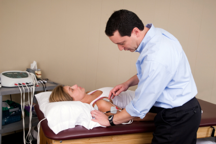
Physical Therapy is a unique rehabilitation technique and art that utilizes a wide variety of procedures such as restoring original functionality and movement to the body, but not limited to eliminating various kinds of pain including Injuries around knee joint, lower back pain, neck pain (cervical) leg pain (sciatica), and post-operative procedures. Typically after being thoroughly evaluated by your physician they generate a specific diagnosis and prescribe physical therapy.

