Tag Archives: Physical Therapy

Patellar Tendinitis


Patellar Tendinitis: Symptoms, Causes and Treatments

The patellar tendon is the one connecting the knee cap to the shin bone. Like other tendons; this tendon as well is made up of hard string like bands. Patellar Tendonitis is an overuse injury resulting from constant strain to patellar tendon.

What do you mean by Patellar Tendinitis?

Patellar Tendinitis also known as Jumper’s Knee is a condition where tendon attaching the patella or knee cap to the top of the shin bone or tibia becomes inflamed, irritated and painful. This condition is more common among the athletes, whose sports involve frequent running, jumping and landing activities like basket ball or volley ball players.

What are the potential signs and symptoms of Patellar Tendinitis?

Pain over the patellar tendon is the initial and noticeable sign of Patellar Tendinitis. Other than this, following symptoms can be noticed in the patients suffering from Patellar Tendinitis:

  • Tender and Swollen tendon
  • Crunching sensation (Crepitus) during the knee movement
  • Feeling pain while jumping or kneeling down
  • Pain while beginning a physical activity
  • Feeling pain while climbing stairs or getting up from a chair
  • Swelling in and around the patellar region
  • Feeling pain while contracting the quadriceps muscles
  • Affected tendon feels thickened than the unaffected one

What are the causes of Patellar Tendinitis?

Patellar Tendinitis is an overuse injury and below mentioned factors contribute towards it:

  • Repeated stress to the patellar tendon
  • Weakening of the tendon structure
  • Repeated stress  to the  supporting structure of knee
  • Inappropriate foot wears
  • Inadequate training techniques
  • Running on the hard surfaces
  • Age
  • Joint laxity and flexibility
  • Misalignment of leg, foot and ankle
  • Flat foot
  • Leg length difference

How Physical Therapy can help to treat Patellar Tendinitis?

Your Physical Therapists will start a proper treatment regime after diagnosing and evaluating your condition properly. The treatment therapies developed usually depend upon the extent and severity of the injury. Therapists may suggest following treatment techniques:

  • Therapists may suggest you to avoid the activities aggravating your condition
  • Eccentric and Concentric program of exercises is designed to treat Patellar Tendinitis. These are the most helpful therapeutic exercises to treat the condition.
  • Splints may be used to immobilize your joint for a short period of time
  • Cold therapy is administered to reduce pain and inflammation
  • A Jumper’s Knee strap may be used by the therapists to lessen the pain and to reduce the pressure on the tendon
  • Stretching exercises may be prescribed to lessen the muscle spam and to lengthen the muscle tendon
  • Strengthening exercises are administered to strengthen the weak thigh muscles
  • Iontophoresis therapy may be used by the therapists
  • Electrical Simulation and Ultrasound may be used to limit and control swelling and pain
  • Flexibility exercises are developed for thigh and calf muscles and to maximize the control and strength of quadriceps muscles
  • Therapists may also design special shoe inserts to improvise the knee alignment and the functioning of patella

Contact Active Physical Therapy for the efficient and state of art treatment of your musculoskeletal problems and disorders. Our certified and acknowledged therapist use patient proven treatment techniques to heal your ailments and to make you as staunch and sturdy as before.
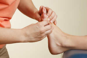
Hammer Toe

Hammer Toe: Causes, Symptoms and Treatment

Hammer Toe

Hammer toe also referred to as Claw Toe is the toe with an inappropriate shape. The toe next to the big toe is the most affected one. This deformity most commonly affects the women wearing high heeled shoes or shoes with narrow toe box.

What do we mean by Hammer Toe?

Hammer Toe is a condition where a bend develops at the joint between the first and second segment of one or more small toes and as a result of which the tip of the toe bends downwards giving it a hammer or claw like shape. Due to the joints of the toe bulging out, the person may have trouble walking. It can turn out to be more serious in the persons with diabetes and poor circulation.

What are the potential causes of Hammer Toe?

Each toe’s muscles work in pairs and whenever there is a muscular imbalance; pressure on the joints and tendons of toe increases and thus toe assumes a hammer head shape. This muscular imbalance occurs due to the following reasons:

  • Poor or ill-fitted shoes including too tight, too short, too pointy and high heeled shoes
  • Foot Structure problem since birth
  • Arthritis
  • Poor blood flow to foot
  • Peripheral Neuropathy (having little or no feeling) in case of diabetic patients
  • Injury to toes
  • Genes also contribute towards developing this condition like flat foot or high arched foot
  • Tight foot ligaments or tendons
  • Spinal cord or peripheral nerve damage

What are the signs and symptoms of Hammer Toe?

Hammer Toe causes you pain and discomfort while walking. Its symptoms may include:

  • A downward bent toe
  • Callus on the sole and corn on the toe of the foot
  • Inability to flex your foot or wiggle your toes
  • Pain while putting a shoe
  • Swelled and red colored toe joint
  • Pain on the ball of the foot
  • Cramping in a toe

What are the treatment options available to treat Hammer Toe?

Treatments are advised according to the severity of your condition. Following treatment options are suggested to treat hammer Toe:

  • Therapists may advice to wear customized toe pads or insoles in your shoes to relieve pain
  • Customized cushions and pads are prescribed to treat bunions and corns
  • Gentle Stretching exercises and Massaging may be recommended to relieve pain and as well to re-position the affected toe.
  • Ice packs are applied to treat painful swelling
  • Exercises are prescribed to restore muscular imbalance
  • Foot exercises are recommended to relax tight foot tendons
  • Manipulation and Splinting of the affected toe is prescribed
  • Exercises to stretch and strengthen the toe muscles are prescribed

Contact Active Physical Therapy for the customized treatment of any of your musculoskeletal deformities and pathologies. Our skilful and dexterous therapists craft out well coordinated and personalized treatment modules to help you to return to your normal activities as early as possible.

Sinus Tarsi Syndrome

Sinus Tarsi Syndrome

Sinus Tarsi Syndrome: Symptoms, Causes and Treatment

Sinus Tarsi Syndrome (STS) is a type of foot pathology, resulting either from the traumatic injury or recurrent injuries or sprain to the ankle during running or walking on a flat foot. It mostly hits athletes or dancers whose professions require a lot of jumping, sudden or quick movements and sudden stops.

 What do we mean by Sinus Tarsi Syndrome (STS)?

The word ‘sinus’ commonly refers to cavity in the bone. The cavity, Sinus Tarsi, is a small cylindrical cavity outside the ankle between the talus and calcaneous bones. Swelling around the Sinus Tarsi region or injury to any of the surrounding ligaments results in Sinus Tarsi Syndrome.

What are the common symptoms associated with Sinus Tarsi Syndrome?

Symptoms related with Sinus Tarsi Syndrome arise gradually over a period of time. Its symptoms are worse during morning but start improving as you warm up. Its symptoms include:

  • Sharp and pinching pain at the top and/or outer side of foot and ankle
  • Pain worsens during the activities like; walking or jumping
  • Stiffness in the ankle
  • Instability is felt while walking or running on uneven ground or slopes and during jumping or changing directions
  • Sinus Tarsi region may be felt tender when touched
  • Pain during ankle movements; especially when you move the sole of foot inwards or downwards
  • Pain intensifies with weight-bearing

What are the causes of Sinus Tarsi Syndrome?

The exact reason of Sinus Tarsi Syndrome is still a matter of debate. Trauma to the ankle is considered to be the most common cause of this pathological condition. Other than this, below mentioned factors give arise to Sinus Tarsi Syndrome:

  • An inversion injury to the foot that is not treated properly
  • Over growth of nerve or fat tissues in the cavity
  • Deviations in bone structures
  • Bone Spurs
  • Arthritis and Bone bridges
  • Ganglion Cysts
  • Foot Deformities (like Flat Foot)
  • Sitting with your feet tucked under you
  • Poor foot Bio-mechanics
  • Inappropriate Footwear
  • Poor flexibility and muscle weakness

How can Sinus Tarsi Syndrome be treated?

Physical Therapy treatment under the guidance and surveillance of expert Physical Therapist is the best treatment option available to treat Sinus Tarsi Syndrome. Therapeutic techniques employed by Physical Therapists not only speed up the recovery but also reduce the chances of recurrence. A complete Physical Therapy Treatment plan consists of:

  • Initially, RICE (Rest, Ice, Compression, Elevation) therapy is advised to to reduce and eliminate pain and to help tissues to heal
  • Activity triggering pain and swelling is identified and eliminated to reduce muscle tension
  • Stable shoes, an ankle sleeve or brace and over the counter or special orthotics are recommended
  • Electrotherapy, laser therapy and cryotherapy is employed

After the pain and swelling are subsided, physical therapists perform following exercises to enable the patient to return to normal activities:

  • Joint mobilisation exercises  are performed to reduce stiffness
  • Stretching exercises including; calf stretches and strengthening exercises are performed
  • Balance Training is provided to prevent  instability
  • Bio-mechanical correction is advised
  • Flexion exercises are administered to strengthen the muscles

 Contact Active Physical Therapy for the state-of-art and effective treatment of any of your musculoskeletal problem and disorders. Our proficient physical therapists create and develop customized treatment plans while taking into view your needs and urgencies.

Turf Toe

What do we mean by Turf Toe?

Turf Toe is the condition referring to the sprain of ligaments around the big toe joint. The injury is referred to as ‘Turf Toe’ as it generally affects the athletes who play on the artificial turf like; football, soccer or baseball players. It is also a common form of injury among the dancers.

But it does not imply that Turf Toe happens to athletes only; anyone whose big toe gets stubbed or jammed suddenly with enough force can develop this condition.

What are the causes leading to Turf Toe?

This injury occurs, when an athlete jams his toe forcefully into the ground or repeatedly pushes it off or bends it backward beyond the normal limits. Apart from these, another contributing factors to Turf Toe Include:

Turf Toe

  • Jamming of the foot on the ground, while rest of the body is pushed forward.
  • Wearing soft-soled foot wears, offering little or no protection to the fore foot.
  • Running and jumping on the hard artificial fields lead to the jamming of big toe and hence to the turf toe.
  • Repeated sprain over the ligaments with enough sudden force can lead to turf toe.
  • Body weight of the person
  • Direct injury that damages the bone under the cartilage.
  • Previous toe or fore foot injuries
  • Positioning of the body while playing sports
  • Yours ankle’s range of motion

What are the symptoms of Turf Toe?

The symptoms of Turf Toe include:

  • Pain at the base of the big toe
  • Tenderness in the ball of foot and big toe
  • Swelling of toe and ball of foot
  • Popping sound can be heard at the time of injury
  • Decreased mobility
  • Bruising of ball of foot and big toe
  • Inefficiency to put weight on the ball of injured foot
  • Failure to push off on the big toe

What are the treatment methods prescribed for Turf Toe?

The best way to treat Turf Toe is to keep the weight or pressure off the toe during the healing process. Treatment methods may include:

  • RICE (Rest, Ice, Compression and Elevation) therapy may be administered to reduce the swelling and pain in the affected area.
  • Athletes are advised to use orthotics or wear stiff soled shoes to secure the toe from more sprain or hyperextension.
  • Strapping or taping of the injured toe to the other toes to provide support and stability.
  • Doctors may immobilize the injured foot in a cast or special walking boot, in order to protect the affected joint

 After a period of immobility and rest, to re-establish the functioning of affected toe; physical therapists practice the following rehabilitation exercises:

  • Passive range of motion exercises are performed to regain function and mobility
  • Flexion and Extension exercises are performed
  • Strengthening exercises like; toe raises, calf raises, squats and lunges are performed
  • Conditioning and Agility exercises are performed

Contact Active Physical Therapy for the proficient treatment of any of your musculoskeletal problems or injuries. Our proficient and eminent physical therapists create customized treatment plans to help you to regain your health and agility.

Treatment for DeQuervain’s Tendonitis

What do you mean by DeQuervain’s Tendonitis?

DeQuervain’s Tendonitis is a painful condition that occurs when the tendons running from the back of your thumb down the side of your wrist get swollen and irritated. This swelling in turn, constricts the canal at the base of your wrist. So, every time when you turn your wrist to hold something or make a fist, it starts aching or hurting.

What are the causes of DeQuervain’s Tendonitis?

DeQuervain’s Tendonitis occurs when you continue with the activities which put strain on your tendons at back / outer aspect of thumb. Factors and activities listed below lead to DeQuervain’s Tendonitis:

de Quervain’s Syndrome.

  • Sports activities like; golf, ten pin bowling, rowing
  • Manual work like; painting, chopping wood, sewing, knitting, use of vibrating machinery
  • Constant lifting of children
  • Direct injury to wrist tendon
  • A fall or forceful movement
  • Injury to thumb, wrist elbow, neck, shoulder and upper back
  • Excessive training
  • Weakness of muscles
  • Muscle Stiffness
  • Tightening of joints
  • Faulty sporting technique
  • Bad posture
  • Poor ergonomic set up
  • Insufficient warm up

 What are the potential signs and symptoms of DeQuervain’s Tendonitis?

Following factors can be stated as potential symptoms of DeQuervain’s Tendonitis:

  • Ache or tightness at the back of thumb or wrist
  • Swollen tendon
  • Feeling of pain when tendon is touched
  • Occasional pins and needles feeling in the thumb
  • Weakness in the wrist, hand and thumb
  • Numbness in the Index finger
  • Movement of thumb and wrist is hindered due to pain and inflammation

How physical Therapy can help to treat DeQuervain’s Tendonitis?

DeQuervain’s Tendonitis is treated using medications, physical or occupational therapy. Discontinuing the activities that affect and worsen your condition, can also help a lot to cure and heal the ailment. Following techniques are employed by the physical therapists to treat DeQuervain’s Tendonitis:

  • Initially, the physical therapist will advice to rest your thumb and wrist to reduce inflammation. At times, splint and braces are used to keep them straight.
  • Manual hands- on therapy is used to treat DeQuervain’s Tendonitis.
  • Soft tissues massage, Trigger point releases, Joint mobilization therapies are administered to improve functioning and relieve strain on tendons.
  • Flexion, Extension and Rotation exercises are performed to strengthen the tendons.
  • Range of motion exercises are administered to loosen and mobilize the stiff tendon.
  • Strengthening exercises are performed to restore functioning and promote healing as well.
  • Stretches are administered to improve the flexibility of the affected tendon.

 At Active Physical Therapy, our eminent and skilled physical therapists are always ready to help you out with any of your musculoskeletal ailments and disorders. Your wellbeing is our top most priority. We are always ready to serve you with best of the treatment plans as per your needs and requirements.


Lumbar Stabilization Program

Lumbar Stabilization: Physical Therapy  Treatment & Management

Lumbar Stabilization is an exercise based program including variety of strengthening and flexibility exercises to heighten a person’s range of motion and as well to strengthen his muscles to support spine. This technique not only improvises his current physical conditions and symptoms but also acknowledge him about efficient movement and safe postures. Lumbar Stabilization program further helps you to find and maintain neutral spine position.

Role of a Physical Therapist

 Before planning and designing a well suited Lumbar Stabilization program, a physical therapist along with your referring physician; thoroughly examines and evaluates you, as the conditions of each individual are unique and specific. Thereafter a multi- dimensional program including strengthening, training, flexibility and endurance exercises is prescribed accordingly to the individual.

Lumbar Stabilization Program

Objectives or Goals of Lumbar Stabilization Program :

  • To attain a neutral spine position
  • To lower or lessen low back pain
  • To cure soft tissue injury like muscle strain
  • To lessen or eliminate lower back injuries that occur due to sudden jerks or movements
  • To exercise control over the forces acting on spine
  • To promote and heighten functional stability

Exercises involved in Lumbar Stabilization Program

During all the exercises performed under Lumbar stabilization program, a neutral spine position is attempted to attain and maintain. As the patient learns to maintain a neutral spine position during current exercise, he is progressed to the next exercise. All the exercises performed under Lumbar Stabilization are patient specific. Following exercises may be recommended under Lumbar Stabilization program :

  • Hamstring Stretch :This is a passive form of exercise, where little muscle effort is used or employed.
  • Pelvic Tilt : An active form of exercise making use of abdominal muscles to move the spine.
  • Arm or Leg raises : A dynamic exercise form, making use of arm or  leg movements while maintaining neutral spine position.
  • Stretching and aerobic exercises are also incorporated in Lumbar Stabilization physical therapy program.
  • Flexibility training is promoted to train muscles and to attain neutral position sine position.
  • Cardiovascular conditioning is combined with Lumbar Stabilization to promote total body strength and endurance.

At Active Physical Therapy, our professional and trained physical therapists ,plan and co-ordinate treatment modules which are designed according to your preferences, capabilities, needs and urgencies. Our treatment modules will not only heal your current ailment but also poses a check on the further ones.

Physical Therapy after a Hip Replacement

Hip Replacement: On the Road to Recovery

When the arthritis in the hip begins to deteriorate and damage the hip joint gradually, hip replacement surgery is performed. But only surgery does not make for the normal and successful functioning of the joint. The battle is won completely when with the help of physical therapy muscles around the hip joint along with the muscles of legs are strengthen and your implanted joint becomes mobilize and begins to work like a normal hip joint.

Hip Replacement Rehabilitation

Need of Hip Replacement Therapy

 Initially after surgery, patients feel stiffness and pain in the hip. So minor Physical Therapy begins on the first day after surgery and is recommended and needed to :

  • Ease and overcome the surgical pain
  • Improve hip’s range of motion
  • Strengthen the muscles around the hip joint
  • Strengthen the muscles of legs
  • Prevent clotting of blood
  • Provide patient education regarding fall, preventions and walking without limp
  • Prevent the loosening and dislocation of the implant

Patient education provided during Hip Replacement Therapy:

  • Patients are advised not to pressurize and stress the hip joint with heavy lifting
  • Specific body posturing and sitting techniques are instructed
  • Patients are briefed not to cross the operated leg over the other one as it can cause the dislocation of the replaced joint
  • Patients are acknowledged about the precautions and safety measures that they should follow while performing the routine activities
  • Patients are educated about the available adaptive  and supportive equipment and their usage

 Exercises recommended under Hip Replacement Therapy:

Physical therapists suggest that to accomplish full recovery and to restore normal hip motion, regular exercises should be performed. During initial days, therapists recommend to exercise 20 to 30 minutes for 2-3 times a day. Following exercises may be suggested and administered by the physical therapists  :

  • Ankle Pumps: It involves pushing your foot up and down.
  • Ankle Rotations: It involves moving your ankle inward and outward the other foot.
  • Buttock Contractions: Involves tightening of buttock muscles.
  • Hip Extension: Involves contracting your hip muscles without holding your breath.
  • Hip Abduction and Adduction: Involves sliding of each leg in and out then back with straight knees and pointed toes.
  • Bridging: Includes lifting up the buttock with bent knees and flat feet.
  • Standing Exercises: These exercises are administered to help you to regain strength for standing independently.
  • Walking, Stair climbing and Descending Exercises: These exercises improvise  strength, mobility and endurance of your knees, hip and legs. Further more, these exercises also help you to regain your balancing skills.

Contact Active Physical Therapy if you are looking forward to the treatment plans that are designed according to your urgencies, preferences and capabilities. Our trained and proficient therapists will provide you with best possible assistance and care and help you to regain your health and well being.

How to cure Lumbar Spondylolysis?

Causes and Symptoms of Spondylolysis:

Lumbar SpondylolysisSpondylolysis happens when a crack forms in the bony ring on the back of the spinal column. Most commonly, this occurs in the low back. In this condition, the bone that protects the spinal cord fractures as a result of excessive or repeated strain. The area affected is called the pars interarticularis, so doctors sometimes refer to this condition as a pars defect.

This condition appears in six percent of children. It mainly affects young athletes who participate in sports in which the spine is repeatedly bent backwards, such as gymnastics, football, and karate.

Anatomy

The human spine is made up of 24 spinal bones, called vertebrae. Vertebrae are stacked on top of one another to create the spinal column. The spinal column gives the body its form. It is the body’s main upright support. The section of spine in the lower back is called the lumbar spine.

Each vertebra is formed by a round block of bone, called a vertebral body. A circle of bone attaches to the back of the vertebra. When the vertebrae are stacked on top of each other, these bony rings create a hollow tube. This tube, called the spinal canal, surrounds the spinal cord as it passes through the spine. Just as the skull protects the brain, the bones of the spinal column protect the spinal cord.

Causes of Lumbar Spondylolysis

Spondylolysis is thought to be caused by repeated strains that damage the lower spine over time. The repeated strains can eventually lead to an overuse injury in the pars interarticularis. The most common location for this to occur is in the lowest vertebra of the spine. This vertebra connects the spine to the pelvis. However, a problem with the pars can occur in any lumbar vertebra. It rarely happens in more than one vertebra at a time.

  • The vertebra initially responds to the abnormal strain by adding new bone cells around the injured area.
  • But if the injuries happen faster than the body can keep up with needed repairs, a crack may form in the weakened bone. This is called a stress fracture.
  • This type of fracture occurs in the pars, the area of bony ring between the pedicle and lamina.
  • The crack may affect only one side of the bony ring. However, it is equally common for the defect to occur on both sides.
  • When this happens, the vertebra is no longer held firmly in place by the facet joints on the back of the ring.
  • As a result, the vertebra is free to slip forward over the one below.
  • This slippage, which is closely related to spondylolysis, is called spondylolisthesis.

Symptoms

  1. People with spondylolysis may feel pain and stiffness in the center of the low back.
  2. Bending fully backward increases pain.
  3. Symptoms typically get worse with activity and go away with rest.
  4. Doctors refer to this type of back pain as mechanical pain because it most likely comes from excess movement between the vertebrae.
  5. Individuals may eventually experience pain that radiates down one or both legs.
  6. This pain may come from pressure and irritation on the nerves that exit the spinal canal near the fracture.
  7. When nerve pressure in the low back causes leg pain, doctors refer it as neurogenic pain.

Treatment

  • Recovery from this condition is much like nonsurgical treatment mentioned earlier.
  • Once you have rested your back to allow it to heal, your doctor may recommend that you work with a physical therapist a few times each week for four to six weeks.
  • In severe cases, patients may need a few additional weeks of physical therapy.

Active’s friendly staff looks forward to assisting you in making your appointment in any of our clinics. Please do not hesitate to contact us, we are dedicated to providing the best physical therapy service in Maryland.

Spinal Cord Injury – Paraplegia

Causes of Spinal Cord Injury:

Spinal Cord InjuryBoth paraplegia and quadriplegia are conditions which result from injury or trauma to the spinal cord. The spinal cord runs through the centre of the bony spine. It is like a system of telephone wires which conduct messages from the brain, through the nerves, to all parts of the body.

Damage can be due to various causes

People can receive damage to the spinal cord due to an accident, for example a:

  • Motor accident
  • Diving mishap
  • Fall
  • Sporting accident
  • Household accident

Damage can also result from a tumour or blood clot on the spinal cord. The majority of people who have a spinal cord injury are young males (who have a greater tendency to indulge in risky behaviour). However, there are people from both sexes and of all ages with spinal cord damage.

Loss of function usually relates to site of Injury

The amount of function lost tends to correspond to the level in the spinal cord where the damage takes place. A break near the top of the neck can mean that even breathing is difficult and that people must use a respirator. People with an injury in the lower neck, however, usually have arm movement but perhaps not finger movement. However they can often drive, work, write etc with the help of specialised equipment.

People with damage to the upper part of the spinal cord are called quadriplegics or tetraplegics. People with damage below this level are called paraplegic.

People with injuries in the lower back can have sensation and movement right to the hips. They can usually live totally independently with a minimum of help, as long as they can use a wheelchair and have the necessary alterations to their home, their bathroom and car.

There are many degrees of injury and function loss, which don’t always correspond to the level of the injury on the spine. Sometimes a quadriplegic only sustains partial damage to the spinal cord and can actually walk, though they may have lost other functions. At all levels, there is usually some loss of function in the bowel and bladder.

Some common Health problems

Other problems which can arise for people with spinal cord injuries are:

  1. Frequent urinary tract infections
  2. Kidney stones
  3. Muscle spasm
  4. Pressure sores from sitting in one position for too long without a correct pressure cushion
  5. Wide and rapid fluctuations in body temperature

Some common causes of Spinal Cord Injury

  • Most spinal cord injuries are caused by accidents.
  • Loss of function tends to correspond to the level in the spinal cord where damage occurred.
  • People with spinal cord injuries can lead productive lives.

Lower back pain occurs due to several causes such as muscle strain, incorrect posture, over-use and spinal injuries, etc. Active Physical Therapy is the best physical therapy clinic in Maryland. Call now for Quick Appointment: 301-662-9335 or Visit http://www.active-physicaltherapy.com

How to Recover from Ankle Sprain?

Causes and Symptoms of Ankle Sprains:

Ankle sprains are a very common ailment. Ligaments are thick, fibrous structures that hold hones together. When these ligaments tear, it’s called a sprain.

Causes of Ankle Sprain

There are multiple causes to sprains, and most are associated with trauma. Running and impact sports put the ankle in precarious positions and can forcibly “roll” or invert the ankle in a painful position.

Symptoms of an Ankle Sprain

  • Ankle sprains are usually associated with pain and swelling and sometimes bruising in the side of the ankle and even into the foot or toes.
  • The symptoms of an ankle sprain vary depending on the severity of the sprain.
  • In most cases, people will experience pain, swelling, instability, and bruising on the inside and outside of the ankle and sometimes down into the foot and toes.
  • Walking can also be painful.

Evaluation of Ankle Sprain

Your physician will examine your ankle to check for signs of a sprain. Also, x-rays are often taken to rule out a fracture.

Prevention of Ankle Sprain

Wearing the proper athletic shoe, supportive brace, taping, stretching, and strength training of the muscles surrounding the ankle joint may assist a person in having a more stable ankle. Following an ankle injury, exercises that promote strength training and balance awareness will significantly reduce one’s likelihood of having a reoccurring ankle sprain.

Common Treatment of Ankle Sprain

  • Immediate treatment should include applying ice to the injured area, with accompanied compression (like an ace wrap) and elevation.
  • The sooner you are able to help control the pain and swelling from the ligament injury, the quicker the rate of recovery and return to function may be.
  • X-rays may be taken to make sure there are no fractures, although these are not always necessary.
  • Activity will be limited as a result of the severity of the injury and the goals of the individual.
  • Crutches, braces and even casting may be required to provide support.
  • The amount of weight bearing is usually determined by how much discomfort there is.

Active Physical Therapy’s experienced dedicated physical therapists and talented clinical team then design individualized treatment plans to achieve the specific goals for each patient per your doctor’s expectation. Call now for Quick Appointment301-916-8540

Visit our Website: http://www.active-physicaltherapy.com