Tag Archives: chronic pain

What is the classification of Shoulder Injury?

Causes and Management of Sternoclavicular Joint:

The Sternoclavicular Joint happens between the proximal end of the clavicle and the clavicular level of the manubrium of the sternum together with a little sector of the first costal fibrous.

Sternoclavicular JointMechanism of Injury

This is the least commonly dislocated joint because of the strong ligaments.

  • Direct force rarely causes this injury. For example, collision of an athlete with another person or a post, etc
  • Indirect force is the most common mode of injury. For example, loading the upper shoulder while someone lies on the sides
  • Incidence is about three percent and is more common in young males.

Causes

Road traffic accident (RTA) is responsible for 80 percent of the cases, sports-related injuries account for the remaining 20 percent.

Classifications

Anatomical classification

Etiological classification

1.       Traumatic

  • Sprain
  • Acute dislocation
  • Recurrent dislocation
  • Unreduced dislocation

2.       Atraumatic

  • Voluntary
  • Involuntary
  • Congenital
  • Degenerative
  • Infective

Clinical Features

The patient complains of pain and swelling. Medial end of the clavicle is prominent in anterior dislocation. Affected shoulder is short. Lateral compression test is positive.

Radiographs

  • AP view is often difficult to interpret.
  • Special 90 degree cephalocaudal views-this helps to see the medial ends of both the clavicles (serendipity view).
  • Tomograms are useful.
  • CT scans and MRI help to study the position of clavicle with respect to sternum and soft tissues respectively.

Management                                                                        

  • Mild sprain: The treatment consists of ice, sling, painkillers, etc.
  • Subluxation: The treatment methods are ice (first 12 hr), warmth (24-48 hr), clavicle strap, and figure of ’8′ and excision of medial end if pain persists.
  • Dislocation: The treatment of choice is closed reduction by firm digital pressure followed by figure of ’8′, clavicle strap, sling, etc. If it fails, open reduction and internal fixation using K-wire is done.

Active Physical Therapy’s experienced dedicated physical therapists and talented clinical team then design individualized treatment plans to achieve the specific goals for each patient per your doctor’s expectation. Call now for best Treatment: 301-877-2323

http://www.active-physicaltherapy.com

Signs and Symptoms of Carpal Tunnel Syndrome

Risk Factors and Prevention of Carpal Tunnel Syndrome:

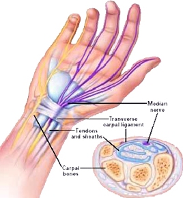
Carpal Tunnel Syndrome (CTS) is a painful and often debilitating disorder of the wrist and hand. The carpal tunnel is a narrow tunnel formed by bones and other tissues on the palm side of your wrist. Within this tunnel runs the median nerve. When the median nerve is compressed or pinched this can lead to CTS. CTS occur when the surrounding tissues in the carpal tunnel, such as ligaments and tendons, get swollen or inflamed and press against the median nerve. This usually results with repetitive motion of the wrist and hand. However, it may also be linked to other factors, such as injury to the wrist such as a fracture.

Carpal Tunnel SyndromeRisk Factors

  • Computer users
  • Grocery checkers
  • Meat packers
  • Violinists
  • Mechanics
  • Pregnant women
  • People diagnosed with diabetes
  • Rheumatoid arthritis
  • Thyroid disease

Diagnosis of Carpal Tunnel Syndrome

CTS are often diagnosed with a detailed history taken either by your doctor or physical therapist. Special tests are then performed to confirm or rule out CTS. If appropriate, a nerve conduction velocity test (NCV) or electromyography (EMG) may be ordered by your doctor to see if the nerves and muscles in your arm and hand show the typical effects of CTS.

Symptoms of Carpal Tunnel Syndrome

  • Numbness and tingling in your hand and fingers (especially the thumb, index and middle finger)
  • Pain in your wrist, palm or forearm
  • Above symptoms are usually greater during the night or with prolonged repetitive motion of the wrist and hand
  • Difficulty gripping objects and weakness in the thumb

Treatment of Carpal Tunnel Syndrome

If CTS is caused by a medical problem your doctor will most likely treat that problem which should help relieve symptoms. Your doctor may also prescribe medication to help decrease inflammation. The anti inflammatory can be administered orally or by way direct injection. Your physical or occupational therapist can also play an integral role by using the appropriate interventions to help loosen the tissue surrounding the median nerve and decrease inflammation.

Prevention of Carpal Tunnel Syndrome

Yes, by utilizing some of the tips below you may decrease your likelihood of developing CTS.

  • Get treatment for any of the diseases listed that predisposes you to CTS
  • Avoid doing the same repetitive movements of the wrist and hand for long periods of time
  • Don’t rest your wrists on hard surfaces for long periods
  • Try to switch hands during work tasks and make sure your tools are appropriate for your hand size
  • Take regular breaks from repeated hand movements

Hand injuries can be caused in many different ways including work injuries, auto injuries, sport injuries, and simply overuse of the muscles causing dysfunction, chronic, and acute pain. We have Certified Hand Therapist in our Laurel and Clinton locations. Call now for Best Hand Therapy Treatment: 301-498-1604 or Visit: http://www.active-physicaltherapy.com

What is Chronic Pain?

Types and Treatment of Chronic Pain:

imagesChronic Pain is commonly considered to signify disease itself. It can be made much more intense by ecological and emotional factors. It continues over a many years than serious discomfort and is immune to most medications. It causes serious problems for sufferers. A person may have two or more co-existing chronic pain conditions.

Types of Pain:

There are two types of pain:

Acute pain doesn’t last long and usually goes away as your body heals. Chronic pain lasts at least 6 months after your body has healed. Sometimes, people who have chronic pain don’t know what is causing it. Along with discomfort, chronic pain can cause low self-esteem, depression and anger. It can also interfere with your daily activities.

Treatment of Chronic pain:

Treatment of chronic pain usually involves medicines and therapy. Medicines used for chronic pain include pain relievers, antidepressants and anticonvulsants. Different types of medicines help people who have different types of pain. You usually use long-acting medicines for constant pain. Short-acting medicines treat pain that comes and goes.

Many other treatments can also relieve pain. They can actually change the body’s chemicals that produce pain. Almost anything we do to relax or get our minds off our problems may help control pain. It’s important to add relaxing activities to your daily life, even if you are already taking medicine for pain. You might have to use stress reduction methods for several weeks before you notice a decrease in pain.

  • Several types of therapy can help ease your pain. Physical therapy (such as stretching and strengthening activities) and low-impact exercise (such as walking, swimming or biking) can help reduce the pain.
  • However, exercising too much or not at all can hurt chronic pain patients. Occupational therapy teaches you how to pace yourself and how to do ordinary tasks differently so you won’t hurt yourself.
  • Behavioral therapy can reduce your pain through methods (such as meditation and yoga) that help you relax.
  • It can also help decrease stress.
  • Lifestyle changes are an important part of treatment for chronic pain.
  • Getting regular sleep at night and not taking daytime naps should help.
  • Stopping smoking helps, also, because the nicotine in cigarettes can make some medicines less effective.
  • Smokers also tend to have more pain than non-smokers.
  • Most pain treatments will not take away all of your pain.
  • Instead, treatment should reduce how much pain you have and how often it occurs.

Active Physical Therapy’s experienced dedicated physical therapists and talented clinical team then design individualized treatment plans to achieve the specific goals for each patient per your doctor’s expectation. Contact now at: 301-916-8540

http://www.active-physicaltherapy.com

How to treat Tibia Bone?

Classification and Treatment of Tibia Bone:

imagesCongenital pseudarthrosis of tibia is a rare condition. It can also be seen in other long bones like femur.

Incidence

  • Incidence is 1 in 2.5 lacs live births.
  • In 50-90 percent of cases neurofibromatosis is present.

Classification

Boyd has classified this condition into six types and is the most accepted classification.

Clinical Features

In this condition, deformity is the chief complaint and the patient develops anterior bowing of the tibia of various severities. In a few cases, there could be pathological fractures.

Radiograph

Radiograph of the leg, AP and lateral views, are sufficient to make an accurate.

Treatment

Principles

  • It depends on the age and type.
  • True pseudarthrosis will not unite with casting alone.
  • Tibial cyst curettage and bone grafting is done in small lesions.
  • Treatment is usually surgical, once a fracture develops.

Physical Therapy is a unique rehabilitation technique and art that utilizes a wide variety of procedures such as restoring original functionality and movement to the body, but not limited to eliminating various kinds of pain including lower back pain, neck pain (cervical), leg pain (sciatica), and post-operative procedures. Call today at: 301-916-8540

http://www.active-physicaltherapy.com

How to get Relief from Knee Injury with Physical Therapy Treatment?

Indications and Treatment of Knee Injury:

Patellofemoral discomfort problem is a common joint issue that causes discomfort under and around the kneecap. The issue often happens because of large exercising that pressures the legs, such as running or moving. In many cases, the discomfort produces from doing too much, too soon. For most people, patellofemoral discomfort decreases with non-surgical treatment, such as anti-inflammatory medicines, physical therapy and rehabilitation.

images

Chondromalacia – Softening and fissuring of the underside of the patella

  • Chondral lesions themselves are asymptomatic unless worn down to subchondral bone.
  • Chondromalacia can only be diagnosed by X-ray (Merchant, sun rise, or skyline view) or surgery.

Presentation

Patellofemoral Syndrome usually presents as an insidious onset of peripatellar or retropatellar pain. Commonly patients are young, active, and females are affected more than males. PFS can also be caused from a traumatic injury to the patella.

Indications

  • Knee pain believed to be musculoskeletal in origin, primarily from muscle imbalances and/or poor biomechanics.
  • Patients report symptoms as general knee pain or ache surrounding the patella.

Treatment

Avoid activities that cause excessive patellofemoral joint reaction forces. Physical Therapy can improve your strength and versatility and avoid the pain from coming back when you continue your regular movement. Your physician or physiotherapist can suggest you on how to progressively return to exercising and how to warm up, awesome down and expand to avoid injury.

Active Physical Therapy is recognized as a provider of superior care for orthopedic, Auto Accident, Sports Injuries / Trauma Cases, Work-related Injuries and comprehensive physical therapy services that improve function, encourage independence and better your quality of life. Call Us at: 301-662-9335

Visit our Website: http://www.active-physicaltherapy.com

What treatment should be done for Hand (Wrist) Injury?

Clinical Features and Investigation of Hand Injury:

imagesTrapezoid bone injuries are the least typical carpal crack of the hand, including less than 1 % of all carpal bone injuries. Its form and place manage security, and dislocation is probably more typical than crack.

Mechanism of Injury

Clinical Features

  • Pain
  • Swelling
  • Tenderness over the wrist
  • Painful resisted flexion are the usual complaints

Investigations

Plain X-rays of the wrist are not reliable. CT scan is a better option.

Treatment

  • Undisplaced fractures are treated by below elbow cast for 4 to 6 weeks.
  • In displaced fractures open reduction and rigid internal fixation is advised.

Physical Therapy is a unique rehabilitation technique and art that utilizes a wide variety of procedures such as restoring original functionality and movement to the body, but not limited to eliminating various kinds of pain. Active Physical Therapy’s experienced dedicated physical therapists and talented clinical team then design individualized treatment plans to achieve the specific goals for each patient per your doctor’s expectation. Call now at: 301-877-2323

http://www.active-physicaltherapy.com

What are the causes of Knee Injury?

Causes and Treatment of Knee Injury:

Osteochondritis dissecans is a combined condition whereby a varying amount of cuboid and its nearby fibrous drops its blood flow. Osteochondritis dissecans can include the cuboid and fibrous of almost any combined. Joints are most commonly affected. In this condition, avascular necrosis occurs in an area of subchondral bone followed usually by degenerative changes in the overlying cartilage. Though it can occur in any joint, it is most commonly seen in the knee joint. This avascular bone undergoes necrosis, gets detached and forms a loose body. Infact, this is the most common cause for loose bodies within the knee.

Knee InjuryCauses

Many causes are cited and are controversial:

  • Exogenous trauma
  • Endogenous trauma
  • Ischemia
  • Abnormal ossification within the epiphysis
  • Genetics
  • Combination of these

Common site

Lateral aspect of the medial femoral condyle near the attachment of posterior cruciate ligament.

Age groups

  • In young patients before epiphyseal closure. Treatment outcome is good.
  • In adults, here treatment outcome is poor.

Clinical Features

  • It is different in the two age groups and consists of vague pain and discomfort in the knee, swelling, catching, popping and locking could occur.
  • After complete separation, loose bodies can be palpated.
  • Tenderness can be elicited over the anteromedial surface of the femoral condyle by deep palpation after flexing the knee.

Investigations

Plain X-rays of the knee (AP, lateral and tunnel view), arthroscopy, bone scan, MRI, etc. are some of the important investigation tools. Plain X-ray also helps to detect the loose bodies of the knee joints.

Treatment

  • This depends on the age of the patient and degree of involvement.
  • Treatment method varies from conservative in children to operative in adults.
  • The operative methods are arthroscopic excision, curettage, pinning, debridement, grafting, etc.
  • The outcome of the treatment is good in children and is not so good in adults.

If you are suffering from Knee Injury then contact today for Quick Appointment at Active Physical Therapy. Active Physical Therapy is the best Physical Therapy Clinic in Maryland.

http://www.active-physicaltherapy.com/

What treatment should be done for Hand Joint Pain?

Treatment of Hand Joint Pain:

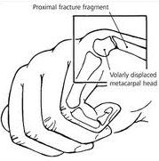
Volar BartonVolar Barton (Palmar rim dislocation) is a palmar rim fracture of distal radius. The most common cause of this type of fracture is a drop on an outstretched hand.

Mechanism

It is due to palmar tensile stress and dorsal shear stress and is usually combined with Radial styloid fracture.

Clinical Features

Radiograph

Palmar rim of distal radial articular surface is displaced dorsally. Proximally and posteriorly and may be associated with dorsal subluxation of the wrist.

Treatment

Conservative

Reduction is simple, but retention is difficult. Long arm cast is used.

Surgery

If reduction does not remain satisfactorily with wrist in neutral or slight palmar flexion, fixation with K-wire, external fixators and buttress plate, etc. may be done. Ellis T-’shaped buttress plate fixation is the preferred method of treatment.

Active Physical Therapy provides state of the art physical therapy throughout the state of Maryland.  Active’s friendly staff looks forward to assisting you in making your appointment in any of our clinics. Contact Us at: 301-498-1604

http://www.active-physicaltherapy.com

How to prevent Hand Disorder?

Types and Treatment of Hand Injury:

indexThis is the fourth most common carpal bone fractures (after scaphoid, capitate and lunate).

Mechanism of Injury

Types of Hand Fracture

These are divided into two types:

  • Body fractures
  • Peripheral chip or avulsion fractures

Clinical Features

Investigations

Plain X-ray of the wrist (AP, lateral or oblique views) may reveal the fracture. If still in doubt, CT scan is recommended.

Treatment

Body Fractures

  • If undisplaced, it is treated by short arm cast for 4 to 6 weeks.
  • If displaced (> 1-2 mm), surgery is the treatment of choice namely:
  • Arthroscopic approach and percutaneus pinning.
  • Open reduction and internal fixation through K-wires or screws.

Chip or Avulsion Fractures

Symptomatic treatment by orthotics in most of the cases helps and rarely may be required in intractable cases.

If you are in Pain and Suffering from Hand Disorder then Visit now at Active Physical Therapy. Please do not hesitate to contact us, we are dedicated to providing the best physical therapy service in Maryland. Call at: 301-877-2323

http://www.active-physicaltherapy.com

How to Remove the disability of Bandy Legs?

Types, Causes and Treatment of Bandy Legs:

images 1Genu Varum  is defined as a lateral angulation of the knee. The longitudinal axis of femur and tibia deviates medially. The deformity involves tibia alone or the femur or tibia  and fibula both.

Types and Causes

Unilateral

  • Due to growth abnormalities of upper tibial epiphysis.
  • Infections like osteomyelitis, etc.
  • Trauma near the growth epiphysis of femur.
  • Tumors affecting the lower end of femur and upper end of tibia.

Bilateral

  • Congenital causes
  • Postural abnormalities
  • Developmental disorders
  • Metabolic disorders (rickets rare)
  • Endocrine disorders
  • Degenerative disorders (e.g.  Osteoarthritis of knee). This is a common cause.
  • Occupational disorders (e.g. in jockeys)
  • Idiopathic
  • Paget’s disease
  • ‘Blounts’ disease (tibia vara)

Clinical Measurements of the Deformity

Child

  • The patient is examined supine with knee extended, patella facing the ceiling and the medial malleoli touching each other. If the separation of the knee exceeds more than 3 cm or if it is unilateral, it should be investigated.
  • A line is drawn from anterosuperior iliac spine through center of patella to medial malleolus. Normally all the structures are in the same line but in genu varum medial malleolus is medial to this line.

Adults

The angle of genu varum is calculated on a standing radiograph of the whole limb.

Clinical Feature

Gena Varum complex: The primary deformity in genu varum is lateral angulation of the knee. In response to this, secondary deformities develop in the tibia and the foot. This together is known as genu varum complex.

Radiograph

Radiograph of the whole limb should be done to assess the severity of genu varum.

Treatment

  • Treatment should be conservative until four years of age. Knee-ankle-foot outhouses with the medial bar and the lateral strap are used.
  • Correction of early deformity is done by dynamic bracing or splints.
  • After four years, significant deformity should be corrected by surgery.
  • Lateral epiphyseal stapling when the child is within the growth period and supracondylar medial open or lateral closed wedge osteotomy is done after skeletal maturity.

Genu varum is a relatively common in kids. Idiopathic shin vara is the most typical of the pathologic circumstances that are associated with bowed legs; therapy techniques differ with the individual’s age and the level of illness and problems. Active Physical Therapy’s experienced dedicated physical therapists and talented clinical team then design individualized treatment plans to achieve the specific goals for each patient per your doctor’s expectation. Call now for Quick Appointment: 301-916-8540

http://www.active-physicaltherapy.com